Trichoscopy for Scalp Disorders: Implementation Protocol
Michele Marchand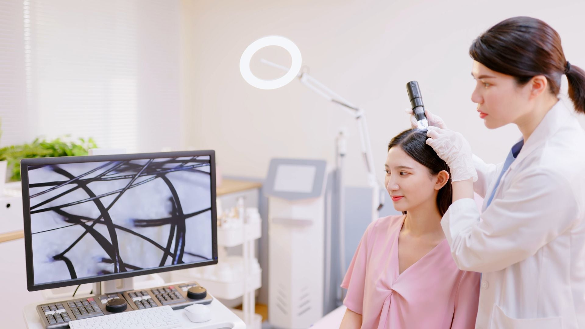
How does trichoscopy help identify and document scalp disorders with accuracy and care?
Table of Contents
- What is trichoscopy and why does it matter for scalp disorders?
- How does trichoscopy work in practice?
- Which scalp disorders can be identified through trichoscopy?
- How is trichoscopy documented and tracked?
- How can trichoscopy guide treatment decisions?
- What should patients expect during a trichoscopy exam?
- How does trichoscopy compare with other diagnostic methods?
- When should someone ask for trichoscopy?
- Final encouragement: Taking the next step with confidence
What is trichoscopy and why does it matter for scalp disorders?
Trichoscopy is a non-invasive diagnostic technique that uses a dermatoscope, a magnifying lens with polarized light, to examine the scalp and hair in fine detail. Unlike a simple visual exam performed in natural light, trichoscopy provides magnified, illuminated views that reveal subtle structural changes invisible to the naked eye. This includes differences in hair shaft texture, abnormalities in follicle openings, or irregularities in the scalp surface. Because it avoids cutting, scraping, or painful interventions, it is considered both safe and comfortable for patients.
For individuals struggling with sensitive scalps, unexplained hair loss, or irritation that does not improve with everyday care, trichoscopy often becomes the first step toward clarity. It reassures patients who feel stuck between confusing product advice and a lack of answers. Trichoscopy is particularly valuable for diagnosing disorders such as psoriasis, seborrheic dermatitis, alopecia areata, and scarring alopecias¹. These conditions often share overlapping symptoms, but their management differs significantly. Accurate identification is key to avoiding unnecessary treatments and preventing long-term scalp damage.
How does trichoscopy work in practice?
A trichoscopy exam is typically performed in a dermatologist’s office using a handheld dermatoscope. The device is placed directly on the scalp surface, sometimes with a drop of mineral oil, alcohol, or gel to enhance image clarity. The magnification can range from 10x up to 70x, allowing clinicians to capture both broad views of affected areas and close-up details of individual follicles or shafts.
Key diagnostic features include:
-
Hair shaft changes: variation in thickness, presence of broken hairs, or twisting and bending patterns.
-
Follicular openings: whether they are intact, clogged with keratin, or absent due to scarring.
-
Vascular patterns: distribution and dilation of small blood vessels that may indicate inflammation.
-
Scaling and pigmentation: presence, color, and texture of flakes, as well as localized discoloration or redness.
Modern clinics often pair trichoscopy with digital photography, creating a permanent record of findings. By comparing results with established trichoscopic image libraries, dermatologists can make accurate, evidence-based diagnoses. For patients, this process feels more like having a specialized photograph taken than undergoing a medical test, which helps reduce anxiety.
Which scalp disorders can be identified through trichoscopy?
Trichoscopy is not limited to one or two conditions. It has become a cornerstone of modern dermatology because of its ability to distinguish between disorders that present with similar outward signs.
-
Alopecia areata (autoimmune hair loss): hallmarked by yellow dots (sebum-filled follicles), black dots (broken hairs at scalp level), and exclamation-mark hairs.
-
Androgenetic alopecia (pattern hair loss): characterized by variability in hair shaft diameters, reduced follicle density, and miniaturization of hair units.
-
Psoriasis of the scalp: identified by thick, silvery-white scales against a red background, often with pinpoint blood vessels.
-
Seborrheic dermatitis: presents with yellowish, greasy scales more diffusely spread, often without significant redness.
-
Lichen planopilaris (scarring alopecia): follicular openings disappear, replaced by scarring tissue, often accompanied by perifollicular scaling and redness.
By linking these visible patterns to specific disorders, dermatologists reduce diagnostic uncertainty. This is especially important for patients with sensitive scalps, who may have tried multiple shampoos, oils, or home remedies without success. With trichoscopy, conditions that appear similar under casual observation can finally be separated with precision.
How is trichoscopy documented and tracked?
Documentation transforms trichoscopy from a single snapshot into a long-term monitoring tool. Dermatologists usually capture high-resolution digital images and enter structured notes into patient records. This not only guides immediate diagnosis but also allows progress tracking across months or years.
The documentation process often includes:
-
Baseline capture: images taken at the first consultation serve as a reference point.
-
Pattern annotation: features such as yellow dots, vascular changes, or scarring are labeled clearly.
-
Comparative follow-up: new images are taken at set intervals, allowing clinicians to see whether inflammation has reduced, shedding has slowed, or regrowth has occurred.
For patients, this visual timeline can be deeply validating. Many people with scalp conditions feel discouraged when progress is slow or invisible day-to-day. Being shown side-by-side images that confirm improvement provides emotional reassurance. For clinicians, standardized documentation supports treatment decisions, enables collaboration with colleagues, and strengthens evidence-based care².
How can trichoscopy guide treatment decisions?
The true power of trichoscopy lies in its ability to connect patterns with specific therapeutic approaches. Because different scalp conditions respond to different interventions, precise diagnosis is essential.
-
Scarring alopecia: If early scarring is detected, urgent anti-inflammatory medications may be started to preserve remaining follicles.
-
Seborrheic dermatitis: Visualization of greasy yellow scales supports treatment with antifungal shampoos, barrier-repair creams, and lifestyle adjustments.
-
Psoriasis: Identification of thick scales and vascular changes may guide use of topical corticosteroids, vitamin D analogs, or systemic therapies for severe cases.
-
Alopecia areata: Detection of black dots and broken hairs can justify initiation of immunomodulatory treatments.
With proper documentation, clinicians can adapt care plans in response to visual evidence. If inflammation subsides on repeat imaging, treatment may be tapered safely. If deterioration is noted, interventions can be escalated before irreversible damage occurs.
What should patients expect during a trichoscopy exam?
For patients who worry about discomfort, it helps to know that trichoscopy is entirely painless. The dermatoscope is simply placed on the scalp, much like a magnifying glass touching the skin. No needles, biopsies, or invasive tools are required in most cases.
What the appointment feels like:
-
Quick: most exams last between 10 and 20 minutes.
-
Safe: no radiation or harmful exposure is involved.
-
Transparent: images are often shared on a screen immediately, allowing patients to see what the dermatologist sees.
-
Collaborative: patients can ask questions during the process, helping them feel more engaged in their care.
Tip for preparation: Bring a list of all hair care products you currently use and note when symptoms first appeared. This context allows your dermatologist to interpret trichoscopic findings in light of your daily habits and history.
How does trichoscopy compare with other diagnostic methods?
While trichoscopy offers detailed visual information, it is not the only diagnostic tool available. Scalp biopsies remain the gold standard when tissue-level confirmation is needed. However, biopsies involve removing a small piece of scalp under local anesthesia, which can be uncomfortable and may leave a small scar.
Trichoscopy, in contrast, is non-invasive and suitable as a first-line tool. It often reduces the need for biopsies by providing clear visual markers that point strongly toward a specific diagnosis. For example, the presence of exclamation-mark hairs in alopecia areata may be sufficient to confirm the condition without tissue sampling.
Compared with simple clinical observation, trichoscopy provides a much higher level of detail. A dermatologist may see redness or scaling with the naked eye, but under trichoscopy, the exact nature of scaling (greasy vs. silvery, thick vs. thin) and vascular distribution becomes clear. This enhanced resolution makes trichoscopy indispensable in modern scalp care³.
When should someone ask for trichoscopy?
Patients often hesitate to request specialized diagnostics, unsure if their concerns are “serious enough.” In reality, trichoscopy is appropriate for many common scenarios:
-
Sudden or patchy hair loss that appears within weeks or months.
-
Persistent itching, burning, or flaking that does not improve with over-the-counter shampoos.
-
A family history of scarring alopecia or premature baldness.
-
Long-standing scalp sensitivity or tenderness without a clear explanation.
Requesting trichoscopy does not mean a condition is severe. Instead, it signals a proactive approach. By catching early signs of disease, trichoscopy can prevent misdiagnosis, reduce wasted time on ineffective treatments, and preserve healthy hair growth whenever possible.
Final encouragement: Taking the next step with confidence
For many people, hair and scalp problems carry more than physical discomfort. They can impact self-image, social confidence, and emotional well-being. It is easy to feel trapped in cycles of self-diagnosis and trial-and-error product testing. Trichoscopy offers an alternative path: clarity, reassurance, and personalized care.
By revealing precise patterns, documenting changes, and guiding evidence-based treatment, trichoscopy strengthens the partnership between patient and dermatologist. If you feel uncertain about what is happening on your scalp, consider discussing trichoscopy at your next appointment. Early intervention brings the best outcomes, and knowing exactly what your scalp is experiencing can restore both comfort and peace of mind.
Glossary
-
Trichoscopy: Non-invasive scalp imaging using a dermatoscope.
-
Dermatoscope: A magnifying device with polarized light for skin and scalp visualization.
-
Alopecia areata: Autoimmune hair loss condition causing patchy bald spots.
-
Androgenetic alopecia: Genetic hair loss, also known as pattern baldness.
-
Psoriasis: Chronic inflammatory skin condition that can affect the scalp.
-
Seborrheic dermatitis: A skin condition causing flaky, greasy patches.
-
Scarring alopecia: Permanent hair loss caused by follicle destruction.
-
Follicular openings: Natural pore structures where hair emerges from the scalp.
-
Perifollicular scaling: Flaky skin around hair follicles.
-
Vascular patterns: Blood vessel changes visible on trichoscopy.
Claims Registry
| Citation # | Claim(s) Supported | Source | Anchor Extract | Notes |
|---|---|---|---|---|
| 1 | "Trichoscopy allows dermatologists to detect subtle structural changes in hair shafts, follicle openings, and scalp skin." | Rudnicka L, Olszewska M, Rakowska A. "Trichoscopy: how it may help the clinician." Dermatol Clin. 2013. | "Trichoscopy enables visualization of hair shafts, follicular openings, and scalp surface features." | Foundational clinical review on trichoscopy. |
| 2 | "Documentation ensures that treatment responses are measurable and evidence-based." | Rakowska A, Slowinska M, Kowalska-Oledzka E. "Trichoscopy in alopecia areata: Clinical utility." J Dermatol Case Rep. 2008. | "Trichoscopy provides a useful method for documenting disease course and treatment efficacy." | Case-based evidence of trichoscopic tracking. |
| 3 | "Compared with simple clinical observation, trichoscopy provides a much higher resolution of details that the naked eye would miss." | Inui S. "Trichoscopy: Hair and scalp dermoscopy." J Dermatol. 2011. | "Trichoscopy reveals diagnostic features not visible to the naked eye." | Widely cited paper establishing trichoscopy as superior to clinical exam. |


