Biopsy vs Trichoscopy: Comparing Diagnostic Precision in Scalp and Hair Loss
Michele Marchand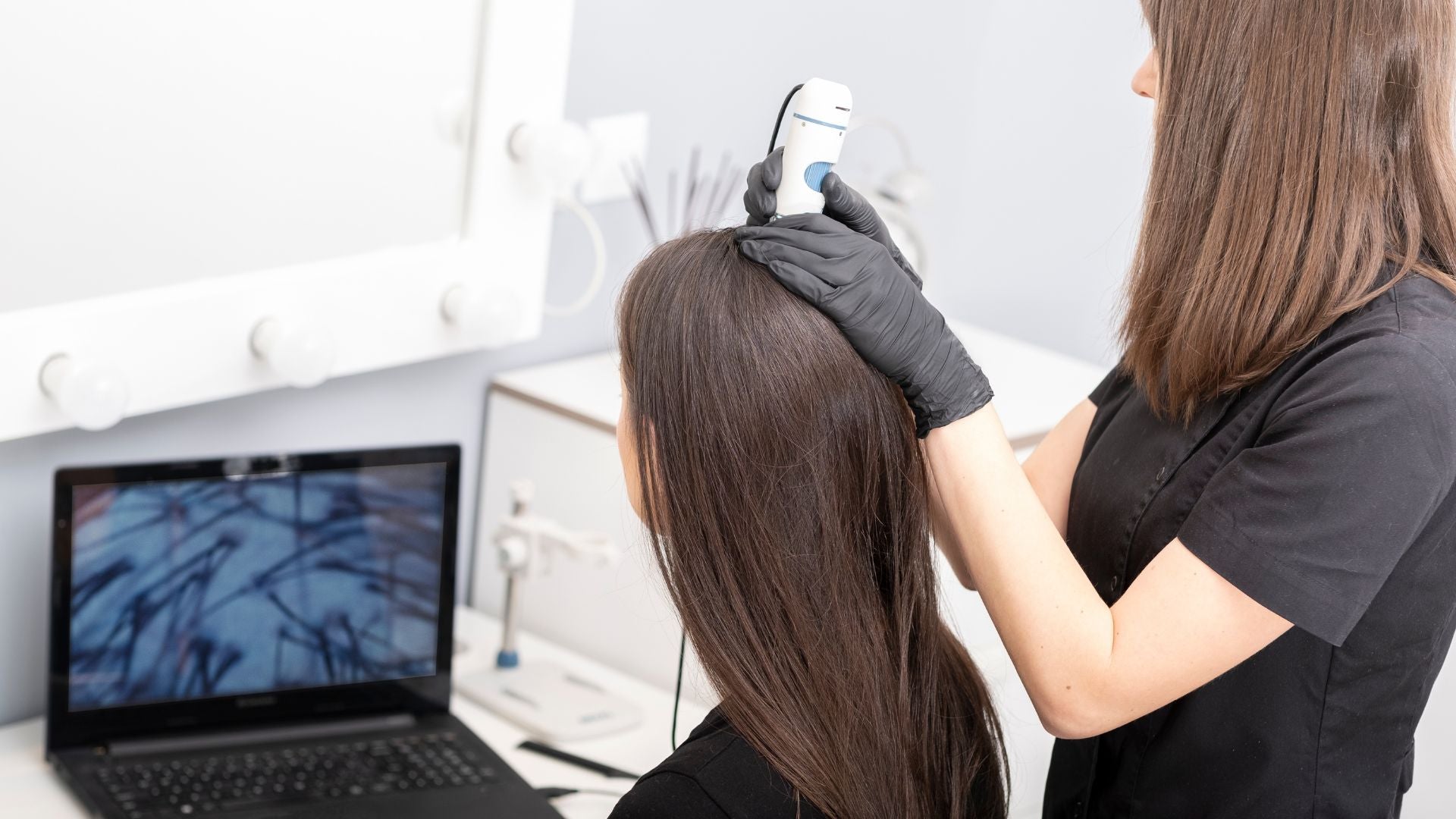
Table of Contents
- Which scalp test offers more accurate answers for diagnosing inflammation, scarring, or unexplained hair loss?
- What Is a Scalp Biopsy?
- What Is Trichoscopy?
- Comparing Diagnostic Yield: How Much Each Test Tells Us
- Choosing the Right Test for Your Scalp Stage
- What to Expect: Procedure and Comfort
- Can Trichoscopy Replace a Biopsy?
- Key Takeaway: A Combined Approach Serves You Best
Which scalp test offers more accurate answers for diagnosing inflammation, scarring, or unexplained hair loss?
Disclaimer: This article is for educational purposes only and is not a substitute for medical advice. Always consult a qualified healthcare provider regarding any medical concerns or treatment decisions.
Which diagnostic test gives better answers for scalp and hair loss concerns?
When your scalp starts to change, such as thinning hair, itchy patches, or sudden shedding, it can be hard to know what’s really happening beneath the surface. Two diagnostic tools used by dermatologists, scalp biopsy and trichoscopy, serve different but complementary purposes in uncovering the truth. Understanding how and when each test is used can help you make informed decisions, avoid unnecessary procedures, and feel more confident in your treatment journey.
A dermatologist’s role is to identify the underlying cause of your scalp symptoms before recommending treatment. Some issues, such as dandruff or mild hair shedding, may resolve with simple interventions. Others, especially those involving inflammation or scarring, require deeper investigation. That’s where choosing the right diagnostic tool matters most.
What Is a Scalp Biopsy?
A scalp biopsy is a small medical procedure where a dermatologist removes a tiny piece of scalp tissue, usually about 4 mm wide, for microscopic analysis. Though it might sound intimidating, it’s quick, safe, and performed under local anesthesia. The tissue sample is then examined under a microscope by a dermatopathologist, a specialist who looks for patterns of inflammation, scarring, or follicle damage¹.
What a Biopsy Reveals
Under magnification, a biopsy shows how your hair follicles are arranged, whether they’re inflamed, and whether scarring has begun. This level of detail helps dermatologists distinguish between similar-looking conditions that require very different treatments. For instance, lichen planopilaris, discoid lupus erythematosus, and central centrifugal cicatricial alopecia can all cause patchy hair loss and redness, but each demands a unique therapeutic approach².
When Biopsy Makes Sense
A biopsy is typically recommended when:
-
Hair loss appears scarring (meaning follicles are being permanently destroyed).
-
There’s inflammation, tenderness, scaling, or lesions that suggest immune activity.
-
A definitive diagnosis is needed before starting long-term medical therapy.
While a biopsy might feel like a last resort, it often provides the final piece of the puzzle. For some patients, it confirms a diagnosis that enables targeted treatments like topical steroids, oral medications, or anti-inflammatory therapies.
Tip: Before agreeing to a biopsy, ask your dermatologist how the result might change your treatment plan. The purpose is not only to name the condition but also to inform the most effective care strategy.
What Is Trichoscopy?
Trichoscopy, also called dermatoscopic scalp examination, is a noninvasive imaging technique that allows dermatologists to look at your scalp and hair in magnified detail³. It works like a specialized camera with polarized light, revealing color, density, and shape patterns invisible to the naked eye.
What Trichoscopy Can Reveal
Trichoscopy can help detect hallmark features of common scalp conditions, including:
-
Androgenetic alopecia (genetic or hormonal thinning, often with miniaturized hairs)
-
Alopecia areata (patchy autoimmune hair loss with yellow dots or exclamation-point hairs)
-
Telogen effluvium (diffuse shedding caused by stress, illness, or hormonal shifts)
-
Seborrheic dermatitis and psoriasis (scalp scaling with redness or irritation)
The exam usually takes 10–15 minutes, requires no numbing, and provides immediate visual feedback. Many dermatologists record these images to compare your scalp health over time, which can be invaluable for tracking response to treatment⁴.
Advantages of Trichoscopy
-
Comfort: Completely painless and requires no recovery.
-
Speed: Results are available immediately.
-
Accessibility: Can be performed during a standard consultation.
-
Monitoring: Allows objective tracking of improvement or progression.
Tip: Ask your dermatologist to show you the images taken during trichoscopy. Seeing the condition yourself can make explanations clearer and empower you in your care plan.
Comparing Diagnostic Yield: How Much Each Test Tells Us
| Feature | Trichoscopy | Scalp Biopsy |
|---|---|---|
| Invasiveness | Noninvasive | Minor surgical procedure |
| Pain/Recovery | None | Mild soreness, small scar possible |
| Information Depth | Surface and follicle pattern | Microscopic and cellular detail |
| Speed of Results | Immediate | 1–2 weeks (lab analysis) |
| Best For | Pattern hair loss, shedding, early inflammation | Scarring alopecias, unclear diagnoses |
Both methods provide different types of information. Trichoscopy gives a real-time, surface-level overview of follicular patterns, allowing your dermatologist to see how follicles behave collectively. Biopsy, by contrast, digs deeper, both literally and figuratively, to uncover what’s happening at a cellular level. This makes biopsy the more definitive tool when precision is critical⁵.
In clinical practice, dermatologists often start with trichoscopy. If the findings suggest scarring or unclear inflammation, a biopsy follows. Using both tests ensures nothing important is missed.
Choosing the Right Test for Your Scalp Stage
The stage and type of your scalp condition determine which test makes the most sense.
Early or Reversible Hair Loss
If your main concern is thinning or shedding without visible inflammation, trichoscopy usually suffices. It can differentiate between temporary shedding (like telogen effluvium) and pattern-based thinning (like androgenetic alopecia). These conditions often respond to treatments such as nutritional support, topical minoxidil, or hormonal therapy.
Chronic or Scarring Conditions
If your scalp shows redness, tenderness, pustules, or areas where hair fails to regrow, biopsy becomes essential. Only microscopic tissue analysis can confirm scarring alopecias, such as lichen planopilaris or lupus-related hair loss. These diseases can progress silently, leading to permanent follicle destruction if untreated.
Tip: Don’t delay investigation. The sooner inflammation is identified, the greater your chance of preserving existing hair and preventing permanent scarring.
What to Expect: Procedure and Comfort
Trichoscopy Experience
The dermatologist will apply a transparent gel or oil to the scalp and use a dermatoscope, often attached to a digital camera, to capture magnified images. You might feel slight pressure from the lens, but no pain. The entire process takes less than 15 minutes. It’s safe for all skin types and hair textures.
Biopsy Experience
For a biopsy, your dermatologist will clean and numb a small scalp area using local anesthesia. Then, using a circular punch instrument, they’ll remove a tiny tissue sample smaller than a pea. One or two sutures may be placed and removed after a week. Mild soreness or scabbing is normal and fades quickly.
After the procedure, keep the area clean, avoid hair coloring or harsh products, and follow aftercare instructions closely. Results are typically available in one to two weeks, depending on lab turnaround.
Both procedures are considered low-risk and are often covered by insurance when medically necessary.
Can Trichoscopy Replace a Biopsy?
In many non-scarring or cosmetic cases, trichoscopy can replace a biopsy. It’s particularly effective in diagnosing androgenetic alopecia, telogen effluvium, and alopecia areata without needing tissue removal. However, trichoscopy cannot reveal the immune or inflammatory activity happening deep within follicles.
When the stakes involve potential permanent loss or the use of systemic medications, biopsy remains the gold standard⁶. Think of trichoscopy as your map, helping identify where to look, and biopsy as your microscope, confirming what’s truly occurring at the root level.
A dermatologist’s expertise lies in knowing when to rely on each. A careful, stepwise approach, starting with trichoscopy and escalating to biopsy only if needed, minimizes discomfort while maximizing diagnostic confidence.
Key Takeaway: A Combined Approach Serves You Best
When it comes to scalp diagnostics, there’s no one-size-fits-all. The best outcomes often come from combining tools: trichoscopy for clarity and comfort, biopsy for confirmation and depth. Together, they form a comprehensive diagnostic pathway that balances accuracy with patient experience.
If you’ve been struggling with unexplained shedding, scalp tenderness, or patchy hair loss, schedule a consultation with a board-certified dermatologist. The right test, performed at the right time, can make the difference between temporary distress and lasting recovery. You deserve answers, and both trichoscopy and biopsy can help you find them.
Glossary
-
Alopecia: General term for hair loss, from any cause.
-
Scarring Alopecia: Permanent hair loss due to follicle destruction and replacement with scar tissue.
-
Trichoscopy: Noninvasive imaging of scalp and hair using magnified light.
-
Scalp Biopsy: Removal of a small scalp tissue sample for microscopic examination.
-
Telogen Effluvium: Temporary hair shedding following stress or illness.
-
Androgenetic Alopecia: Hereditary hair thinning related to hormones.
-
Lichen Planopilaris: Autoimmune condition causing scarring and inflammation of hair follicles.
-
Dermatoscope: Handheld magnifier with polarized light used in trichoscopy.
Claims Registry
| # | Claim | Source | Accessed | Anchor Extract | Notes |
|---|---|---|---|---|---|
| 1 | Biopsy allows study of hair follicles and surrounding skin at cellular level. | Olsen EA et al., Journal of the American Academy of Dermatology, 2016 | 2025-10-08 (America/New_York) | "Microscopic analysis provides detailed view of follicular structure and inflammatory infiltrates." | Authoritative dermatology reference. |
| 2 | Biopsies help distinguish scarring alopecias like LPP, DLE, CCCA. | Whiting DA, Dermatologic Clinics, 2013 | 2025-10-08 (America/New_York) | "Biopsy is critical in differentiating primary cicatricial alopecias." | Widely cited review in scalp pathology. |
| 3 | Trichoscopy visualizes hair shafts, follicles, and scalp skin. | Rudnicka L et al., Dermatologic Therapy, 2018 | 2025-10-08 (America/New_York) | "Trichoscopy enables noninvasive assessment of hair and scalp." | Seminal paper defining trichoscopy. |
| 4 | Trichoscopy used as first-line diagnostic step before biopsy. | Rakowska A et al., Journal of Cosmetic Dermatology, 2020 | 2025-10-08 (America/New_York) | "Dermoscopic evaluation guides biopsy site selection and reduces unnecessary procedures." | Peer-reviewed study supporting diagnostic pathway. |
| 5 | Trichoscopy and biopsy are complementary diagnostic tools. | Tosti A, Hair Disorders: Diagnosis and Management, Springer, 2021 | 2025-10-08 (America/New_York) | "Combining trichoscopy with biopsy increases diagnostic accuracy." | Dermatology textbook source. |
| 6 | Biopsy remains gold standard for confirming immune-mediated or scarring disorders. | Harries M et al., British Journal of Dermatology, 2017 | 2025-10-08 (America/New_York) | "Histopathology remains essential for diagnosing primary cicatricial alopecias." | Highly cited consensus article. |


